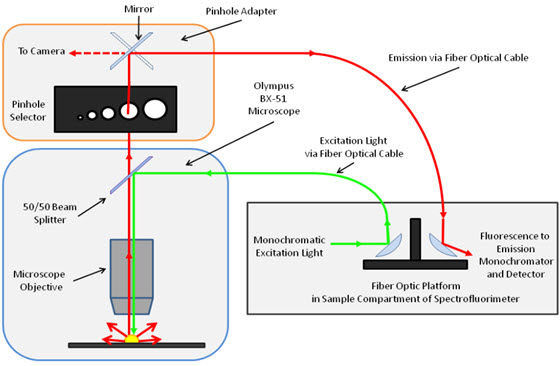
Although fluorescence microscopy is commonly used in forensic labs to examine fibers, the researchers in this project said that by removing specific signal filters from their instruments they have “taken the non-destructive nature of fluorescence microscopy to a higher level of selectivity.” Some forensic practitioners do not take advantage of the full potential of fluorescence microscopy for the analysis of textile fibers, they said, but by acquiring excitation-emission matrixes (EEM) that are typically screened out with “band-pass filters, the researchers gained valuable identification information in single fibers.
When those filters are in place, the researcher said, forensic investigators are unable to record excitation and emission spectra from their fluorescence microscopes and, as a result, limit the information they can collect on the spectral features of textile fibers. “The instrumentation presented [in this study] removes this limitation by coupling a microscope to a spectrofluorimeter.” See Figure 1.
The NIJ-supported research, conducted by scientists at The University of Central Florida, looked not only at impurities imbedded into fibers during the fabrication of garments, but also at the spectral changes that can occur in textile fibers as a result of exposure to environmental conditions such as laundering, cigarette smoke, and weathering. The researchers collected visible absorption spectra from three pairs of dyed fabrics, with each pair containing dyes with highly similar chemical structures.
Beyond identifying impurities in textile fibers, the researchers were able to determine if a fiber had been washed and noted that, “characterization of fibers based on the detergents used in laundering could be a powerful extension of this work and would potentially enable identification of a detergent used on textiles without solvent extraction or destruction of the fiber.”
The effects of weathering on the spectral signals from fibers varied with the types of fibers. The researchers called for future studies to compare fibers from humid versus desert environments.
The scientists also looked at what traces cigarette smoke left on various cloth materials. Fibers on the surface of the cloth and directly exposed to cigarette smoke were compared with fibers deeper in the fabric. The deeper fibers showed no exposure to the smoke and a week after exposure even the surface fibers showed only negligible evidence of cigarette smoke.
The researchers concluded that, by conducting the fluorescence microscopy without the EEM filters they have taken fiber analysis to a “higher level of selectivity.” Because of that increased selectivity, they were able to determine that intrinsic fluorescence impurities imbedded into the fibers is a ‘reproducible source of fiber comparison.”
About This Article
The research described in this article was funded by NIJ cooperative agreement number 2011-DN-BX-K553, awarded to the University of Central Florida. This article is based on the grantee report "Comparison of Microspectrophotometry and Room-Temperature Fluorescence Excitation-Emission Matrix Spectroscopy for Non-Destructive Forensic Fiber Examination" (pdf, 21 pages) by Andres D. Campiglia, University of Central Florida.


