Aviso de archivo
Esta es una página de archivo que ya no se actualiza. Puede contener información desactualizada y es posible que los enlaces ya no funcionen como se pretendía originalmente.
Home | Glossary | Resources | Help | Contact Us | Course Map
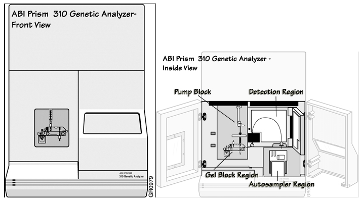
The most commonly used capillary electrophoresis (CE) units are manufactured by Applied Biosystems. All Applied Biosystems CE instruments operate similarly. The two most common models used in forensic science are the ABI 310 and the ABI 3100. This section focuses on the ABI Prism® 310 Genetic Analyzer instrument. Other CE instruments used in forensic DNA laboratories are listed at the end of this module.
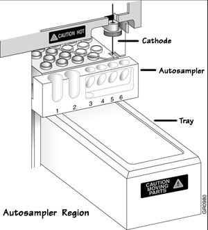
Autosampler Region | |
|---|---|
| Component | Purpose |
| Autosampler | Holds the sample tray and tubes and presents them to the capillary for injection |
| Cathode Electrode | Provides a negative pole for electrical current and is composed of platinum |
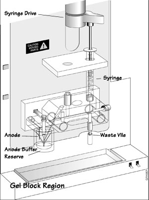
Gel Block Region | |
|---|---|
| Component | Purpose |
| Syringe | Stores the polymer between runs and generates the necessary force to fill the capillary with polymer |
| Anode Electrode | Provides a positive electrical current and is composed of platinum |
| Anode Buffer Reservoir | Contains buffer required for electrophoresis |
| Syringe Drive | Provides positive pressure to the syringe |
| Waste Vial | Collects waste |
Ions migrate through the capillary during electrophoresis. Positive ions will gather at the cathode and negatively charged ions will gather at the anode. The movement of ions creates an imbalance called buffer depletion. Buffer depletion can impair separation of DNA fragments due to a reduction in current. It is important to replenish or replace the buffer regularly to compensate for buffer depletion.
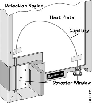
Detection Region | |
|---|---|
| Component | Purpose |
| Charge-Coupled Device (CCD) | Detects fluorescence after the laser excites the dyes in the sample. The CCD camera is located behind the detector window. |
| Heat Plate | Heats the capillary during electrophoresis |
| Capillary | Where separation occurs. The capillary is a hollow fused silica tube with an internal diameter of 50-100µm and 25-75cm in length. There is an outer polyimide coating that provides physical strength while allowing a degree of flexing of the silica tube. The capillary is filled with polymer and is transparent to ultraviolet and visible light. The capillary itself can be used as the detector cell when fluorescence detection is used. Before each injection, the capillary is filled with a new aliquot of a viscous polymer solution. |
| Detection Cell Window | Where dye tagged product is detected. The detection cell window is produced by burning the polyimide outer coating off the capillary. This cleared area is directly in the line of the laser beam to detector path, allowing the DNA fragments to be visualized as they pass by. Note: The detection cell window makes the capillary fragile in this area. |
Computer | |
|---|---|
| Component | Purpose |
| Computer | Controls the operation of the instrument. The instrument settings are defined in method files in the computer. These settings consist of run temperature, voltage, injection parameters, rinse cycles, and sample positions. |
Additional Online Courses
- What Every First Responding Officer Should Know About DNA Evidence
- Collecting DNA Evidence at Property Crime Scenes
- DNA – A Prosecutor’s Practice Notebook
- Crime Scene and DNA Basics
- Laboratory Safety Programs
- DNA Amplification
- Population Genetics and Statistics
- Non-STR DNA Markers: SNPs, Y-STRs, LCN and mtDNA
- Firearms Examiner Training
- Forensic DNA Education for Law Enforcement Decisionmakers
- What Every Investigator and Evidence Technician Should Know About DNA Evidence
- Principles of Forensic DNA for Officers of the Court
- Law 101: Legal Guide for the Forensic Expert
- Laboratory Orientation and Testing of Body Fluids and Tissues
- DNA Extraction and Quantitation
- STR Data Analysis and Interpretation
- Communication Skills, Report Writing, and Courtroom Testimony
- Español for Law Enforcement
- Amplified DNA Product Separation for Forensic Analysts


