Aviso de archivo
Esta es una página de archivo que ya no se actualiza. Puede contener información desactualizada y es posible que los enlaces ya no funcionen como se pretendía originalmente.
Home | Glossary | Resources | Help | Contact Us | Course Map
Sample Preparation
Samples run on Capillary electrophoresis (CE) instruments are amplified as described in course: DNA Amplification, the Multiplexing Module before placing them into the autosampler tray.
Electrokinetic Injection
There are three methods used for sample introduction in CE systems:
- Hydrodynamic or pressure injection
- Siphoning
- Electrokinetic injection05
All of these methods require the immersion of the capillary end into the sample. Electrokinetic injection is the only method used in forensic DNA analysis.
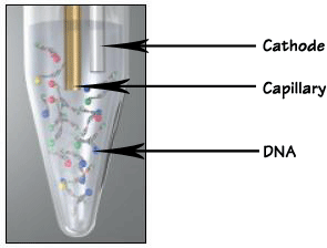
The DNA sample is loaded into the capillary separation matrix by electrokinetic injection. The process consists of the transfer of negatively charged ions via an electromotive force. As current flows from the cathode to the anode, the DNA sample is introduced at the cathode end of the capillary. Because only negatively-charged ions are transferred from the sample in the process, there is no significant loss of sample volume.
This type of transfer is directly influenced by the ionic strength of the sample.06 The magnitude and duration of the voltage applied to the capillary during electrokinetic injection is directly proportional to the amount of DNA loaded into the capillary. Ideally, only the DNA present in the sample will contribute to the ionic strength. The presence of competing ions other than DNA rapidly degrades or prevents the injection process.
Electrokinetic injections are highly efficient in separating the DNA fragments due to the sample stacking in the capillary. Sample stacking in the capillary results when samples are injected from a solution that has a lower ionic strength than the buffer inside the capillary. When the electric field is applied during an electrokinetic injection, the resistance and field strength in the sample plug region increase because there are fewer ions to carry the current in the lower ionic strength samples. This causes the ions from the sample to migrate rapidly into the capillary. When these sample ions enter a region where the polymer solution and buffer are at higher ionic strength, they slow down and stack as a sharp band at the boundary between the sample plug and the electrophoresis buffer (Butler 1995).
Additional Online Courses
- What Every First Responding Officer Should Know About DNA Evidence
- Collecting DNA Evidence at Property Crime Scenes
- DNA – A Prosecutor’s Practice Notebook
- Crime Scene and DNA Basics
- Laboratory Safety Programs
- DNA Amplification
- Population Genetics and Statistics
- Non-STR DNA Markers: SNPs, Y-STRs, LCN and mtDNA
- Firearms Examiner Training
- Forensic DNA Education for Law Enforcement Decisionmakers
- What Every Investigator and Evidence Technician Should Know About DNA Evidence
- Principles of Forensic DNA for Officers of the Court
- Law 101: Legal Guide for the Forensic Expert
- Laboratory Orientation and Testing of Body Fluids and Tissues
- DNA Extraction and Quantitation
- STR Data Analysis and Interpretation
- Communication Skills, Report Writing, and Courtroom Testimony
- Español for Law Enforcement
- Amplified DNA Product Separation for Forensic Analysts


