Home | Glossary | Resources | Help | Contact Us | Course Map
Aviso de archivo
Esta es una página de archivo que ya no se actualiza. Puede contener información desactualizada y es posible que los enlaces ya no funcionen como se pretendía originalmente.
Reverse Lighting Technique
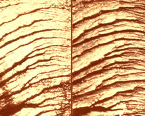
To document fractured comparisons, the objects should be viewed side-by-side on separate stages of a comparison microscope. The fractured surface of each specimen can be made to appear as a mirror image of the other on each side of the optical hairline through the use of the reverse lighting technique. The results are very graphic and convincing in demonstrating that the two objects were once joined together, especially after image documentation is generated.
The following steps are a guide for conducting an examination using reverse lighting:
- Orient both items such that they appear to be in their original position relative to each other.
- Place the items on a laboratory bench running from left to right.
- Make an index mark (using a permanent marker) from one corresponding surface to the other, crossing over the interface where the fracture apparently occurred. For instance, if a fractured rod appears to have once been joined, place an indexing mark on the two sections so that the index mark crosses over the fracture line from left to right.
- Hold the temporarily joined specimens so that the indexing line produced by the permanent marker is vertical and facing toward the examiner.
- Rotate the top end of the upper specimen 180 degrees and embed the nonevidentiary end in a specimen holder (clay, plasticine, etc.) on the left stage of the microscope with the indexing mark facing toward the examiner.
- Embed the bottom end of the lower specimen in a specimen holder on the right stage of the microscope with the indexing mark facing toward the examiner.
The end result of the placement of the specimens on their respective stages will be two questioned fractured objects prepared for comparison using reverse lighting. - Arrange the microscope lights for grazing illumination, one arranged in the conventional way (directed toward the examiner from the back of the microscope) and one in an unconventional way (mounted in the front of the stage directed away from the examiner).
- Bring both items into focus at low illumination for a side-by-side comparison.
- Note that although the two surfaces are different (high points on one are low points on the other), the shadows generated give the appearance of two mirror images on either side of the optical hairline because one of the lights is reversed by 180 degrees.
- The area of best agreement should be documented, preferably by digital or conventional photography or by other means, as determined by laboratory protocol. Images should be marked with the examiners initials, case identifier, degree of magnification, item numbers, and a description of the nature of the observed area.
- Examine both items for the presence of manufacturing marks. Manufacturing marks provide corroborative information that can further support the fracture identification. These marks should also be documented by some type of imaging.
- Examine the alignment of the broken edges of both items at ninety degrees to the fractured surface for additional corroboration. Imaging should be used to support the overall conclusion.
Fracture matches (physical matches) typically represent a small percentage of the toolmark caseload, but may be encountered in some cases, such as in terrorism incidents involving fractured bomb components.
Click here to read a sample forensic worksheet and report - Toolmark Identification (Fractures)
Selected Bibliography
The Selected Bibliography is a list of the writings that have been used in the assemblage of the training program and is not a complete record of all the works and sources consulted. It is a compilation of the substance and range of readings and extensive experience of the subject matter experts.
- AFTE Criteria for Identification Committee. 1992. Theory of identification, range striae comparison reports and modified glossary definitions AFTE criteria for identification committee report. AFTE J 24 (2): 336-340.
- AFTE. 1998. Theory of identification as it relates to toolmarks. AFTE J 30 (1): 86.
- Biasotti, A., and J. Murdock. 1984. Criteria for identification in firearms and toolmark identification. AFTE J 16 (4): 16-24.
- Biasotti, A., and J.E. Murdock. 1997. Firearms and toolmark identification: Legal issues and scientific status. In Modern Scientific Evidence: The Law and Science of Expert Testimony , ed D.L. Faigman, D.H. Kay, M.J. Saks, and J. Sanders, 124 151. St Paul: West Publishing Co.
- Brackett, J. 1970. A study of idealized striated marks and their comparisons using models. J of Forensic Sci Soc 10 (1): 27-56.
- Butcher, S., and D. Pugh. 1975. A study of marks made by bolt cutters. J of Forensic Sci Soc 15, (2): 115-126.
- Collins, R., and R.S. Stone. 2005. How unique are impressed toolmarks? An empirical study of 20 worn hammer faces. AFTE J 37 (4): 252-295.
- Davis, J.E. 1958. Introduction to Tool Marks, Firearms and the Striagraph . Springfield: Charles C Thomas Pub Ltd.
- Ernest, R. 1991. Toolmarks in cartilage revisited. AFTE J 23 (4): 958-959.
- Galan, J. 1986. Identification of a knife wound in bone. AFTE J 18 (4): 72-75.
- Hall, J. 1992. Consecutive cuts by bolt cutters and their effect on identification. AFTE J 24 (3): 260-272.
- Hatcher, J. 1935. Textbook of Firearms Investigation, Identification and Evidence . Plantersville: Small-Arms Technical Publishing Co.
- Hatcher, J. 1947. Hatchers Notebook . Harrisburg: Military Service Publishing Co.
- Hatcher, J.S, F.J. Jury, and J. Weller. 1957. Firearms Investigation, Identification, and Evidence . Harrisburg: Stackpole Books.
- Kockel, R. 1980. About the appearance of clues or marks from knife blades. AFTE J 12 (3): 16-28.
- Locke, R. 2006. Characteristics of knife cuts in tires. AFTE J 38 (1): 56-65.
- May, L. 1930. The identification of knives, tools, and instruments: A positive science. Am J of Police Sci 1 (3): 246-259.
- Miller, J., and M. McLean. 1998. Criteria for identification of toolmarks. AFTE J 30 (1): 15-61.
- Miller, J. 2000. Criteria for identification of toolmarks Part II. AFTE J 32 (2): 116-131.
- Miller, J., and G. Beach. 2005. Toolmarks: Examining the possibility of subclass characteristics. AFTE J 37 (4): 296-345.
- Moran, B. 2003. Photo documentation of toolmark identification An argument in support. AFTE J 35 (2): 174-189.
- Nichols, R. 2003. Consecutive matching striations (CMS): Its definition, study and application in the discipline of firearms and toolmark identification. AFTE J 35 (3): 298-306.
- Stone, R. 2003. How unique are impressed toolmarks? AFTE J 35 (4): 376-383.
- Thompson, E., and R. Wyant. 2003. Knife identification project (KIP). AFTE J 35 (4): 366-370.
- Tomasetti, K. 2002. Analysis of the essential aspects of striated toolmark examination and the methods for identification. AFTE J 34 (3): 289-301.
- Walsh, K., and G. Weavers. Jan March 2003. Toolmark Identification: Can We Determine a Criteria? INTERface Forensic Sci Soc News Letter 29
- Watson, D. 1979. The identification of toolmarks from consecutively manufactured knife blades in soft plastic. AFTE J 10 (3): 43-45.
Additional Online Courses
- What Every First Responding Officer Should Know About DNA Evidence
- Collecting DNA Evidence at Property Crime Scenes
- DNA – A Prosecutor’s Practice Notebook
- Crime Scene and DNA Basics
- Laboratory Safety Programs
- DNA Amplification
- Population Genetics and Statistics
- Non-STR DNA Markers: SNPs, Y-STRs, LCN and mtDNA
- Firearms Examiner Training
- Forensic DNA Education for Law Enforcement Decisionmakers
- What Every Investigator and Evidence Technician Should Know About DNA Evidence
- Principles of Forensic DNA for Officers of the Court
- Law 101: Legal Guide for the Forensic Expert
- Laboratory Orientation and Testing of Body Fluids and Tissues
- DNA Extraction and Quantitation
- STR Data Analysis and Interpretation
- Communication Skills, Report Writing, and Courtroom Testimony
- Español for Law Enforcement
- Amplified DNA Product Separation for Forensic Analysts


