Home | Glossary | Resources | Help | Contact Us | Course Map
Aviso de archivo
Esta es una página de archivo que ya no se actualiza. Puede contener información desactualizada y es posible que los enlaces ya no funcionen como se pretendía originalmente.
Stereo and Comparison Microscopes
Comparison microscopy is the most important technique in the field of forensic firearms/toolmark examination and comparison. Similar to other forms of visible light microscopy, it is enhanced to allow simultaneous examination of two separate objects. These microscopes allow the simultaneous comparison of the unique striations and impressed marks found on the surfaces of objects bearing toolmarks (including fired bullets and cartridge cases).
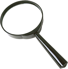
The most primitive form of microscopy uses a convex lens to make objects appear larger. These lenses are normally convex on both sides and have a short focal length. Whenever an object is placed within this short focal length, an image is produced that is erect and larger than the original object. An excellent modern example of this is the simple handheld magnifier. The magnifying power of primitive lenses was not very high. Nonetheless, it was a significant accomplishment that paved the way for the next evolutionary step the compound microscope.
| Note: |
| As far back as 2600 B.C. the Egyptians noticed the phenomenon of magnification using rock crystals with a roughly convex shape. Later, the Greeks and Romans practiced glass blowing and observed the magnifying effect of water in curved glass containers. They also used convex lenses to focus and magnify the heat from the sun. The Arab mathematician Alhazen described how the lens of the human eye forms an image on the retina. In mid-thirteenth century England, Roger Bacon referred to the use of convex lenses in many of his works. During the same period, frame spectacles came into use. |
Compound Microscopy
Credit for the invention of the compound microscope is often given to Zacharias Janssen, a spectacle maker from Middleburg, Holland (1590s). In 1610, Galileo announced his version, followed by many other prominent pioneers in the field of optics. They found that when two simple convex lenses with specific characteristics were placed at the ends of a tube with a defined distance between them, much greater magnifications could be achieved. This constituted the first compound microscope one containing more than a single lens. The lower lens (nearest to the object being observed) was called the objective lens; the lens closest to the observer was called the eyepiece or ocular lens. When the entire assembly was raised or lowered, the object being observed could be brought into focus. When the object is brought into clear focus, an inverted image is formed by the objective lens at a point within the principle focus of the ocular lens. In turn, this image becomes the object for the ocular lens. This results in an even larger image for the observer. These early compound microscopes typically had magnifications of 3x to 9x.
This was a great leap forward, but there were optical problems with both single lens and compound microscopy.
The problems included
- dimensional distortion,
- color inaccuracies (especially at the edges of the round convex lenses used in the systems),
- inverted images.
These and other problems were corrected by the use of additional combinations of complex concave and convex lenses added to the microscope lens tube. By 1873, German Ernst Leitz had invented the revolving nosepiece for quickly rotating objective lenses.
Stereomicroscopy
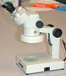
Stereomicroscopy is based on the use of physically joined compound microscopes (one for each eye) in the examination of single objects. Sometimes a single objective lens is shared by both eyepieces. This approach yields a three-dimensional (stereo) view of an object, such as a fired bullet being evaluated prior to microscopic comparison with another bullet. It is particularly useful when combined with an optical zoom feature, as is common in many stereomicroscopes. The stereomicroscope is useful during any phase of forensic firearms work, including the next level of visible light microscopy comparison microscopy.
Maintenance and Calibration
The Association of Firearm and Tool Mark Examiners (AFTE) provides its members with a set of standard techniques for the maintenance and calibration of stereomicroscopes. While these techniques are not mandatory (unless stated in individual laboratory policy), they are widely used to satisfy the requirements of accreditation standards.
The standards for preparing a stereomicroscope for actual case work are as follows:
- Annually:
- The stereomicroscope will be cleaned, serviced and certified (by a factory certified technician).
- These steps will be documented in the instruments maintenance/calibration logbook.
- Quarterly:
- The stereomicroscope will be standardized utilizing a 100-0.01 division 3x objective. This objective will either be obtained from the manufacturer or have a NIST traceable certificate.
- The microscope may be calibrated with a ruler with divisions of 0.01. This ruler should also have a NIST traceable certificate.
- This will also be documented in the instruments maintenance/calibration logbook.
- For each use:
- The stereomicroscope will be checked to ensure that it is functioning properly.
- This check will be performed by observing an item under the microscope and utilizing past experience to determine if the instrument appears to be giving a true and accurate representation of the evidence.
- This check does not need to be documented.
Additional Online Courses
- What Every First Responding Officer Should Know About DNA Evidence
- Collecting DNA Evidence at Property Crime Scenes
- DNA – A Prosecutor’s Practice Notebook
- Crime Scene and DNA Basics
- Laboratory Safety Programs
- DNA Amplification
- Population Genetics and Statistics
- Non-STR DNA Markers: SNPs, Y-STRs, LCN and mtDNA
- Firearms Examiner Training
- Forensic DNA Education for Law Enforcement Decisionmakers
- What Every Investigator and Evidence Technician Should Know About DNA Evidence
- Principles of Forensic DNA for Officers of the Court
- Law 101: Legal Guide for the Forensic Expert
- Laboratory Orientation and Testing of Body Fluids and Tissues
- DNA Extraction and Quantitation
- STR Data Analysis and Interpretation
- Communication Skills, Report Writing, and Courtroom Testimony
- Español for Law Enforcement
- Amplified DNA Product Separation for Forensic Analysts


