Aviso de archivo
Esta es una página de archivo que ya no se actualiza. Puede contener información desactualizada y es posible que los enlaces ya no funcionen como se pretendía originalmente.
Home | Glossary | Resources | Help | Contact Us | Course Map
Some of the proteins circulating in serum display detectable polymorphisms, with alleles that have sufficient frequency differences to be of value in blood typing. Transferrin (Tf) and Group Specific Component (Gc) were two that offered considerable promise and were becoming routinely used just before the advent of DNA typing. However, haptoglobin (Hp) was the most widely used of the polymorphic serum proteins in forensic biology.
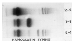
Haptoglobin is a hemoglobin-binding protein found in the α-globulin fraction of serum. There are two alleles, designated Hp 1 and Hp 2, with several rare variants at each allele. The alleles are separated by electrophoresis on a gradient polyacrylamide gel (that is, one in which the concentration of polyacrylamide varies from 5% at the top to 30% at the bottom, so giving enhanced separation by molecular sieving).
Haptoglobin 1 is a monomer consisting of two pairs of peptide chains (α and β) joined by disulfide bridges. Electrophoresis of serum from someone who is homozygous for Hp 1 shows only one band. In contrast, samples from someone who is homozygous for Hp 2 display multiple bands on electrophoresis. Curiously, electrophoresis of a sample from a heterozygous Hp 2-1 shows a band matching the Hp 1 band along with multiple other bands, but these do not align exactly with those from a haptoglobin 2 homozygous person. The Hp 2 proteins are similar to Hp 1 in that they are composed of α and β peptide chains cross-linked by disulfide bridges. However, the α peptides (α2) are not the same as those in Hp 1. Furthermore, the proteins are found as polymers of the structure α2nβn where n is between 3 and 8. In heterozygotes, some of the polymers incorporate α1 chains as well as α2 ones.03, 04,
Haptoglobin is a reasonably good system for use in forensic serology. It is stable in stains and the assay is quite sensitive, using one of the hemoglobin screening procedures, such as leucomalachite green, to visualize the bands by reacting with the bound hemoglobin.
Additional Online Courses
- What Every First Responding Officer Should Know About DNA Evidence
- Collecting DNA Evidence at Property Crime Scenes
- DNA – A Prosecutor’s Practice Notebook
- Crime Scene and DNA Basics
- Laboratory Safety Programs
- DNA Amplification
- Population Genetics and Statistics
- Non-STR DNA Markers: SNPs, Y-STRs, LCN and mtDNA
- Firearms Examiner Training
- Forensic DNA Education for Law Enforcement Decisionmakers
- What Every Investigator and Evidence Technician Should Know About DNA Evidence
- Principles of Forensic DNA for Officers of the Court
- Law 101: Legal Guide for the Forensic Expert
- Laboratory Orientation and Testing of Body Fluids and Tissues
- DNA Extraction and Quantitation
- STR Data Analysis and Interpretation
- Communication Skills, Report Writing, and Courtroom Testimony
- Español for Law Enforcement
- Amplified DNA Product Separation for Forensic Analysts


